





A brain MRI is a detailed imaging test that uses magnetic fields to create high-resolution pictures of the brain. It is one of the most important tools for diagnosing neurologic conditions.
Quick Facts:
- Provides highly detailed images of the brain
- Does not use radiation
- Commonly used for tumors, stroke, and neurologic symptoms
- Often performed with or without contrast
👉 Request an Appointment
👉 Refer a Patient
WHAT IS A BRAIN MRI?
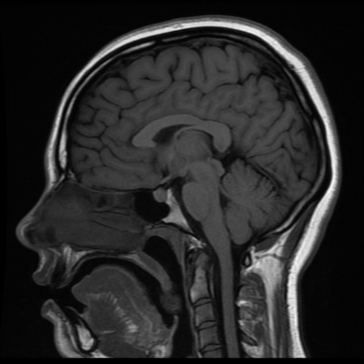
A brain MRI (magnetic resonance imaging) uses a powerful magnet and radio waves to produce detailed images of the brain.
It can show:
- Brain tissue in high detail
- Small abnormalities not visible on CT
- Changes in blood flow and fluid spaces
- Tumors, inflammation, and structural conditions
Unlike CT scans, MRI:
- Does not use radiation
- Provides greater detail for soft tissues
WHY IS A BRAIN MRI PERFORMED?
A brain MRI is used to evaluate a wide range of neurologic conditions.
Common Reasons for a Brain MRI
- Headaches with concerning features
- Brain tumors or masses
- Stroke or neurologic symptoms
- Seizures
- Weakness, numbness, or balance problems
- Evaluation of known conditions (e.g., Chiari, hydrocephalus)
👉 The goal is to identify and characterize brain abnormalities with precision
WHAT CAN A BRAIN MRI DETECT?
MRI is one of the most sensitive imaging tests for the brain.
Common Findings
- Brain tumors (benign and malignant)
- Stroke (especially early or small strokes)
- Chiari malformation
- Hydrocephalus
- Multiple sclerosis and inflammation
- Vascular abnormalities (with specialized sequences)
When MRI Is Preferred Over CT
- Evaluating tumors or subtle lesions
- Assessing chronic or progressive symptoms
- Detailed imaging of brain structures
TYPES OF BRAIN MRI
MRI Without Contrast
- Provides structural detail
- Used for many initial evaluations
MRI With Contrast
- Uses a contrast agent (gadolinium)
- Highlights tumors, inflammation, and blood vessels
- Helps distinguish different types of abnormalities
Specialized MRI Techniques
- Diffusion imaging (stroke detection)
- Functional imaging (in select cases)
- MR angiography (blood vessels)
HOW THE TEST IS PERFORMED
Step-by-Step Overview
- Preparation
- You lie on a table that slides into the MRI scanner
- Imaging
- The scanner uses magnetic fields to create images
- The machine makes loud noises during scanning
- Contrast (if needed)
- Contrast dye may be given through an IV
- Completion
- The test typically takes 20–45 minutes
OUR APPROACH
We use brain MRI as part of a thoughtful, diagnosis-driven evaluation.
- MRI is often the most informative imaging test for brain conditions
- We interpret imaging in the context of symptoms and clinical findings
- We use MRI to guide decisions about observation, further testing, or treatment
- The goal is to provide clarity and avoid unnecessary procedures
👉 Imaging is a tool—clinical judgment determines what it means
WHAT TO EXPECT
Before the MRI
- You may be asked about metal implants or devices
- Most MRIs require no special preparation
During the MRI
- The test is painless
- You will need to lie still
- Some patients may feel confined (open MRI or sedation may be options)
After the MRI
- Most patients resume normal activity immediately
- Results are reviewed by your physician
OUTCOMES & BENEFITS
Potential Benefits
- Highly detailed imaging of brain structures
- Early detection of disease
- Improved diagnosis and treatment planning
- No radiation exposure
MRI is essential for:
- Accurate diagnosis
- Long-term monitoring
- Surgical planning
RISKS & CONSIDERATIONS
MRI is very safe, but considerations include:
- Not suitable for some metal implants or devices
- Claustrophobia in some patients
- Rare reactions to contrast dye
- Longer scan time compared to CT
👉 Most patients tolerate MRI very well
FREQUENTLY ASKED QUESTIONS
Is MRI better than CT?
It depends. MRI provides more detail, while CT is faster and better for emergencies.
Do I need contrast?
Not always. It depends on what your doctor is looking for.
Is MRI safe?
Yes, it is very safe and does not use radiation.
What if I’m claustrophobic?
Options include open MRI or mild sedation.
FOR REFERRING PHYSICIANS (Optional Expandable Section)
- Indications for MRI brain:
- Tumor evaluation
- Chronic or progressive neurologic symptoms
- Seizures
- Suspected structural abnormalities
- MRI with and without contrast preferred for:
- Tumors
- Infection
- Inflammatory conditions
- Consider additional imaging:
- MRA/MRV
- CTA
- Follow-up MRI for monitoring
RELATED CONDITIONS / PROCEDURES
- Brain Tumors
- Chiari Malformation
- Hydrocephalus
- Stroke & Intracranial Hemorrhage
- Head CT