





A catheter angiogram is a detailed imaging procedure used to visualize blood vessels in the brain. It is considered the most accurate test for diagnosing conditions such as aneurysms, vascular malformations, and certain types of stroke.
Quick Facts:
- Gold standard for imaging brain blood vessels
- Provides highly detailed, real-time images
- Performed using a small catheter inserted through an artery
- Often used when precise vascular detail is needed
👉 Request an Appointment
👉 Refer a Patient
WHAT IS A CATHETER ANGIOGRAM?
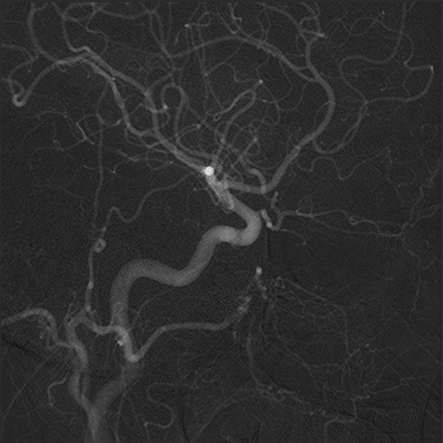
A catheter angiogram is a minimally invasive procedure that uses X-ray imaging and contrast dye to visualize blood vessels in the brain.
During the procedure:
- A thin catheter is inserted into an artery (usually in the wrist or groin)
- The catheter is guided to blood vessels in the neck or brain
- Contrast dye is injected to make the vessels visible on imaging
This produces detailed, real-time images of blood flow and vascular anatomy.
WHY IS THIS PROCEDURE PERFORMED?
A catheter angiogram is used when detailed evaluation of brain blood vessels is required.
Common Reasons for a Catheter Angiogram
- Brain aneurysm evaluation
- Arteriovenous malformation (AVM)
- Subarachnoid hemorrhage
- Stroke or vascular narrowing
- Evaluation of abnormal findings on CT or MRI
👉 The goal is to accurately diagnose vascular conditions and guide treatment decisions
WHAT MAKES THIS DIFFERENT FROM CTA OR MRA?
While CT angiography (CTA) and MR angiography (MRA) are non-invasive, catheter angiography provides:
- Higher resolution imaging
- Real-time visualization of blood flow
- Ability to evaluate small or complex vessels
- Option for immediate treatment in some cases
👉 It is often used when precision is critical
WHO IS A CANDIDATE?
Patients may be candidates if:
- Non-invasive imaging is inconclusive
- A vascular condition requires detailed evaluation
- Treatment planning requires precise anatomy
- There is suspected aneurysm, AVM, or vessel abnormality
HOW THE PROCEDURE IS PERFORMED
Step-by-Step Overview
- Preparation
- The procedure is typically performed in a specialized imaging suite
- Anesthesia
- Local anesthesia with sedation is commonly used
- Catheter Insertion
- A small catheter is placed into an artery (wrist or groin)
- Navigation
- The catheter is guided to blood vessels supplying the brain
- Contrast Injection
- Dye is injected while X-ray images are taken
- Completion
- The catheter is removed and pressure is applied to the insertion site
OUR APPROACH
We use catheter angiography as part of a precise and individualized diagnostic strategy.
- Not all patients require invasive angiography
- We use it when detailed vascular information will change management
- Imaging findings are integrated with clinical evaluation
- The goal is to guide the safest and most effective treatment plan
👉 Precision in diagnosis leads to better outcomes
WHAT TO EXPECT
Before the Procedure
- Review of imaging and medical history
- Instructions regarding medications and fasting
During the Procedure
- Typically lasts 30–90 minutes
- Performed with sedation
- Patients are awake but comfortable
After the Procedure
- Observation for several hours
- Limited activity for the remainder of the day
- Most patients go home the same day
OUTCOMES & BENEFITS
Potential Benefits
- Highly accurate diagnosis of vascular conditions
- Better treatment planning
- Ability to detect small or complex abnormalities
- Real-time assessment of blood flow
Catheter angiography is often essential for:
- Definitive diagnosis
- Surgical or endovascular planning
RISKS & COMPLICATIONS
As with any invasive procedure, risks include:
- Bleeding or bruising at the catheter site
- Stroke (rare)
- Allergic reaction to contrast dye
- Vessel injury (rare)
👉 These risks are low but are carefully considered before recommending the procedure
FREQUENTLY ASKED QUESTIONS
Is this the same as a CT angiogram?
No. CT angiography is non-invasive, while catheter angiography is more detailed and invasive.
Will I be asleep?
Usually not. Most patients receive sedation but remain comfortable.
Is it painful?
Discomfort is usually minimal, with local anesthesia used at the insertion site.
Can this treat the problem as well?
In some cases, treatment can be performed during the same session, depending on the condition.
FOR REFERRING PHYSICIANS (Optional Expandable Section)
- Refer patients with:
- Suspected or known aneurysm
- AVM or vascular malformation
- Subarachnoid hemorrhage
- Inconclusive CTA/MRA
- Imaging:
- CTA or MRA often performed first
- Angiography for definitive evaluation
- Consider referral for:
- Treatment planning
- Complex vascular anatomy
- Multidisciplinary evaluation
RELATED CONDITIONS / PROCEDURES
- Brain Aneurysm
- Vascular Malformations (AVMs)
- Stroke & Intracranial Hemorrhage
- CT Angiography (CTA)
- MR Angiography (MRA)