





A head CT (computed tomography) scan is a fast and widely used imaging test that creates detailed pictures of the brain. It is often the first test used to evaluate head injuries, stroke, or sudden neurologic symptoms.
Quick Facts:
- Quick and widely available imaging test
- Often used in emergency situations
- Excellent for detecting bleeding and fractures
- Typically completed in just a few minutes
👉 Request an Appointment
👉 Refer a Patient
WHAT IS A HEAD CT?
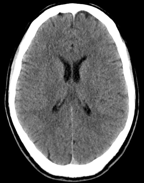
A head CT scan uses X-rays and computer processing to create detailed images of the brain and skull.
It can show:
- Bleeding in or around the brain
- Skull fractures
- Swelling or mass effect
- Changes related to stroke
Unlike MRI, CT scans are:
- Faster
- More readily available in emergency settings
- Often the first imaging test performed
WHY IS A HEAD CT PERFORMED?
A head CT is commonly used to quickly evaluate serious neurologic conditions.
Common Reasons for a Head CT
- Head injury or trauma
- Suspected stroke
- Severe or sudden headache
- Confusion or altered mental status
- Seizures
- Weakness, numbness, or speech difficulty
👉 The goal is to quickly identify urgent problems that require treatment
WHAT CAN A HEAD CT DETECT?
A head CT is especially useful for identifying:
Common Findings
- Intracranial hemorrhage (bleeding)
- Subdural or epidural hematoma
- Skull fractures
- Hydrocephalus (fluid buildup)
- Large brain tumors or masses
Limitations
- May not detect very small lesions
- Less detailed than MRI for certain conditions
- Early stroke changes may not always be visible
👉 CT is often the first step, with MRI used for further detail if needed
HOW IS THE TEST PERFORMED?
Step-by-Step Overview
- Positioning
- You lie on a table that slides into the scanner
- Imaging
- The scanner rotates around your head
- Images are captured in seconds
- Completion
- The scan usually takes only a few minutes
In some cases:
- Contrast dye may be used to enhance certain structures
OUR APPROACH
We use head CT as part of a rapid and accurate diagnostic process.
- CT is often the first imaging step in urgent situations
- We carefully interpret imaging in the context of symptoms and exam findings
- When needed, we recommend additional imaging (such as MRI or angiography)
- The goal is to quickly identify problems and guide appropriate treatment
👉 Imaging is one part of a complete clinical evaluation
WHAT TO EXPECT
Before the Scan
- Usually no special preparation is needed
- You may be asked about allergies if contrast is used
During the Scan
- The test is painless
- You will need to lie still briefly
After the Scan
- Most patients can return to normal activity immediately
- Results are reviewed by a physician
OUTCOMES & BENEFITS
Potential Benefits
- Rapid diagnosis of serious conditions
- Widely available in emergency settings
- Helps guide immediate treatment decisions
- Non-invasive and quick
Head CT is often critical for:
- Emergency decision-making
- Early detection of life-threatening conditions
RISKS & CONSIDERATIONS
Head CT is generally safe, but considerations include:
- Exposure to a small amount of radiation
- Rare reactions to contrast dye (if used)
- May require additional imaging for full evaluation
👉 The benefits of CT imaging often outweigh risks in urgent situations
FREQUENTLY ASKED QUESTIONS
Is a head CT the same as an MRI?
No. CT is faster and better for emergencies, while MRI provides more detailed images.
Do I need contrast?
Not always. Many CT scans are done without contrast.
How long does it take?
Usually just a few minutes.
Is it safe?
Yes. It is widely used and considered safe when medically appropriate.
FOR REFERRING PHYSICIANS (Optional Expandable Section)
- Indications for CT head:
- Acute neurologic symptoms
- Head trauma
- Suspected hemorrhage
- CT is preferred initial imaging for:
- Stroke evaluation
- Trauma
- Altered mental status
- Follow-up imaging may include:
- MRI brain
- CTA/MRA
- Repeat CT if condition changes
RELATED CONDITIONS / PROCEDURES
- Head Trauma (TBI)
- Stroke & Intracranial Hemorrhage
- Hydrocephalus
- Brain Tumors
- MRI Brain