





Advanced Imaging of the Spine
A spine MRI provides detailed images of the bones, discs, nerves, and soft tissues of the spine.
It is one of the most important tools used to diagnose the source of neck, back, arm, or leg symptoms and helps guide treatment planning.
👉 Request an Appointment
👉 Refer a Patient
What Is a Spine MRI?
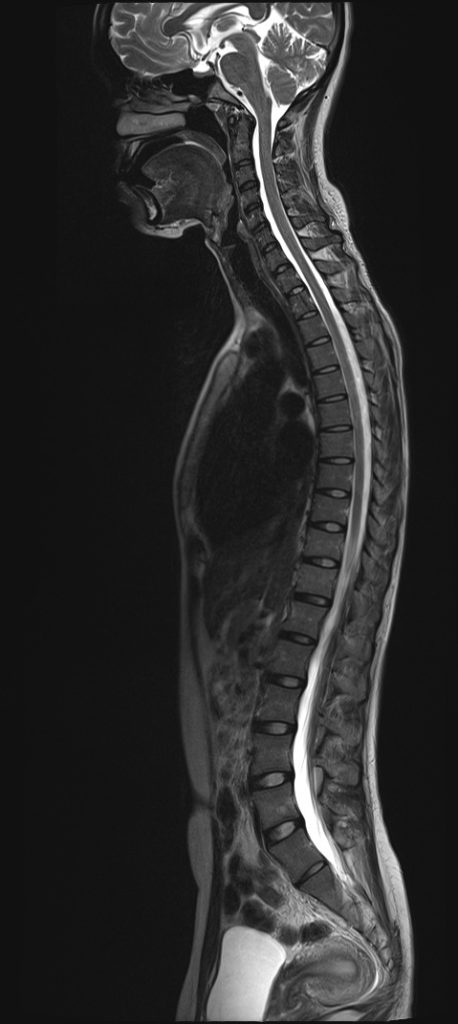
MRI stands for Magnetic Resonance Imaging.
This test uses a powerful magnet and radio waves to create highly detailed images of the spine without radiation exposure.
A spine MRI allows your physician to evaluate:
- Spinal discs
- Nerve roots
- Spinal cord
- Vertebrae
- Ligaments and soft tissues
Why Might I Need a Spine MRI?
Your physician may recommend an MRI if you have:
- Neck or back pain
- Arm or leg pain
- Numbness or tingling
- Weakness
- Balance problems
- Persistent symptoms that have not improved
MRI is especially helpful when symptoms suggest nerve compression or spinal cord involvement.
What Conditions Can It Help Diagnose?
A spine MRI can identify:
- Herniated discs
- Spinal stenosis
- Degenerative disc disease
- Nerve compression
- Spinal cord compression
- Tumors
- Infections
- Fractures
- Inflammation
What to Expect During the Test
During the MRI:
- You will lie on a padded table
- The table slides into the scanner
- You must remain still while images are taken
- The machine makes loud tapping noises (ear protection is provided)
The scan typically takes 30–60 minutes.
Some studies may require contrast dye for additional detail.
Why MRI Matters in Spine Care
MRI helps your physician:
✓ Identify the cause of symptoms
✓ Determine the exact location of nerve compression
✓ Guide treatment decisions
✓ Plan surgery when needed
It is one of the most valuable tools for creating an accurate diagnosis.
Is MRI Safe?
MRI is very safe for most patients.
Be sure to tell your care team if you have:
- A pacemaker
- Metal implants
- Claustrophobia
- Prior implanted medical devices
Alternative imaging options may be recommended if needed.